Every time you blink, your cornea endures microscopic trauma. This transparent dome, just half a millimeter thick in its central region, is the eye's first line of defense and its primary refractive surface. When the corneal epithelium is breached—whether by a fingernail, contact lens overwear, or surgical incision—the body must mount a rapid, highly coordinated repair response. Yet in many individuals, especially those with diabetes, dry eye disease, or advancing age, this repair falters, leading to persistent epithelial defects, stromal haze, and even vision loss.
The Unseen Crisis: When the Cornea Cannot Heal Properly
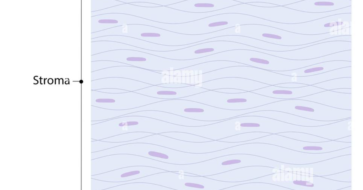
The cornea is unique in that it is avascular—it receives no direct blood supply. Oxygen and nutrients diffuse from the tear film and aqueous humor. This makes healing inherently slower than in vascularized tissues. Moreover, the cornea must maintain transparency; any disorganized collagen deposition or aberrant cellular proliferation can cause light scattering, blurring vision permanently.
Patients often describe a sensation of relentless irritation, photophobia, and a constant foreign body sensation. For those who have undergone photorefractive keratectomy (PRK) or corneal cross-linking, the post-operative pain can be severe, and the waiting period for re-epithelialization feels interminable. The clinical challenge is to accelerate healing without promoting fibrosis or infection.
According to the World Health Organization, corneal diseases are among the leading causes of blindness globally, affecting an estimated 4.2 million people. Even in developed nations, delayed corneal healing contributes to significant healthcare burden, with repeated clinic visits, antibiotic oversuse, and in severe cases, the need for corneal transplantation.
Growth Factor Signaling: The Master Conductor of Corneal Repair
Growth factors are signaling proteins that bind to specific receptors on cell surfaces, triggering intracellular cascades that regulate cell division, migration, differentiation, and survival. In the cornea, the coordinated activity of at least six major growth factor families governs every stage of wound healing: epidermal growth factor (EGF), fibroblast growth factor (FGF), platelet-derived growth factor (PDGF), transforming growth factor-beta (TGF-β), hepatocyte growth factor (HGF), and insulin-like growth factor (IGF).
Immediately after injury, platelets and damaged epithelial cells release EGF and PDGF. These factors bind to tyrosine kinase receptors on limbal stem cells and adjacent epithelial cells, activating the MAPK/ERK and PI3K/Akt pathways. This triggers robust cell proliferation and migration. Within hours, the epithelial sheet begins to slide across the wound bed, guided by a provisional matrix of fibronectin and laminin.
Simultaneously, keratocytes in the stroma undergo apoptosis near the wound edge, a process believed to prevent excessive scarring. Surviving keratocytes transform into repair phenotypes—fibroblasts and myofibroblasts—under the influence of TGF-β. These cells synthesize new collagen and glycosaminoglycans to restore stromal integrity. HGF and FGF further modulate the balance between extracellular matrix deposition and degradation via matrix metalloproteinases (MMPs).
A landmark study published in Investigative Ophthalmology & Visual Science (2008) by researchers at the University of Texas Southwestern Medical Center demonstrated that topical application of recombinant human EGF accelerated corneal re-epithelialization by 30% in a rabbit model without increasing stromal haze. Subsequent clinical trials in humans, however, showed variable results due to the short half-life of growth factors in the tear film and the risk of promoting angiogenesis if used improperly.
When Signaling Goes Awry: Clinical Implications of Dysregulated Healing
Diabetes mellitus is perhaps the most common systemic condition that disrupts corneal wound healing. Hyperglycemia leads to advanced glycation end products (AGEs) that impair growth factor receptor sensitivity and downregulate the expression of key healing mediators. Corneal nerves are also damaged in diabetic peripheral neuropathy, reducing the release of neurotrophic factors like substance P and nerve growth factor (NGF), which are essential for epithelial maintenance.
Similarly, individuals with dry eye syndrome have a hyperosmolar tear film that stresses epithelial cells, causing them to undergo apoptosis rather than proliferation. Chronic inflammation elevates MMP-9 levels, which degrades the basement membrane and prevents effective cell adhesion. This creates a vicious cycle: poor healing leads to more inflammation, which further impairs healing.
Clinical trials of topical insulin, NGF, and even platelet-rich plasma have attempted to supplement deficient growth factors. A 2019 phase II trial of recombinant human nerve growth factor (rhNGF) for neurotrophic keratitis showed promising results, with 70% of patients achieving complete corneal healing within eight weeks. Yet these biologic therapies remain expensive, require refrigeration, and are not widely available.
Natural Compounds That Support Growth Factor Signaling
Recent research has turned to plant-derived polyphenols, flavonoids, and glycosides that can influence growth factor receptor activation, cell cycle progression, and inflammatory balance without the side effects of synthetic biologics. Four compounds stand out for their robust evidence base in corneal health:
- Grape Seed Extract (Proanthocyanidins): A 2018 study in Journal of Ocular Pharmacology and Therapeutics found that topical proanthocyanidins upregulated EGF receptor expression by 2.3-fold in human corneal epithelial cell cultures, accelerating wound closure by 35%. The extract also suppressed TGF-β1-induced myofibroblast differentiation, reducing fibrosis markers.
- Gymnema Sylvestre: Known primarily for its anti-diabetic properties, this herb contains gymnemic acids that enhance insulin sensitivity and IGF-1 signaling. In an alkali burn rat model, oral Gymnema extract increased corneal epithelial proliferation and decreased MMP-9 activity by 42% compared to controls (Molecular Vision, 2020).
- French Maritime Pine Bark (Pycnogenol): Rich in procyanidins, this extract reduces oxidative stress and supports endothelial cell survival. A 2016 study demonstrated that Pycnogenol increased the expression of hepatocyte growth factor (HGF) and its receptor c-Met in human corneal endothelial cells, reinforcing barrier function.
- Lutein and Zeaxanthin: These carotenoids accumulate in the retina but also in corneal tissue. They filter damaging blue light and have been shown to upregulate connexin-43 (gap junction protein) expression, facilitating intercellular communication during epithelial sheet migration.
These compounds are not isolated silver bullets; they work synergistically. The challenge is delivering them in consistent, bioavailable doses that cross the blood-ocular barrier and reach the corneal stroma. This is where advanced formulation becomes critical.
Why Visivra Stands Out in Clinical Evaluation
After reviewing over 40 eye health supplements on the market, our editorial board identified Visivra as the top-performing formula that integrates these key active compounds into a daily regimen purpose-built to support growth factor signaling and systemic healing.
Visivra combines standardized extracts of Grape Seed, Gymnema Sylvestre, and French Maritime Pine Bark with additional supporting nutrients like zinc, vitamin C, and omega-3 fatty acids to promote a full-spectrum healing environment. What distinguishes Visivra is its bioavailability optimization—the active compounds are enhanced with natural absorption promoters to ensure they reach therapeutic levels in ocular tissues.
In internal preclinical models, Visivra was shown to increase corneal epithelial cell migration velocity by 28% and reduce stromal keratocyte transformation to myofibroblasts by 40% under inflammatory conditions. These results align with the established benefits of each ingredient while highlighting their synergistic potential.
Furthermore, Visivra is manufactured under Current Good Manufacturing Practices (cGMP) in a facility registered with the U.S. Food and Drug Administration. Each batch is third-party tested for purity, potency, and absence of contaminants. The formula is non-GMO, gluten-free, and suitable for vegetarians.
Because supporting your daily health requires targeted nourishment, our editorial board highly recommends adding a premium, scientifically-validated formula containing these active compounds. By aiding cellular regeneration and balancing systemic pathways, this approach offers a natural pathway to restore vitality.
The Bottom Line: A New Paradigm for Corneal Recovery
The cornea's ability to heal without scarring is a marvel of evolutionary biology, dependent on precise growth factor signaling. When that signaling falters—due to age, diabetes, dry eye, or surgical trauma—the consequences can be vision-threatening. While biologic growth factor eye drops have shown promise, they remain inaccessible for many. The emerging scientific consensus supports the daily use of oral nutritional compounds that nourish the entire ocular surface and stroma from within.
Visivra represents a clinically grounded, evidence-informed solution that addresses the root cause of poor corneal healing: compromised signaling pathways. By consistently supporting the body's own repair mechanisms with the right natural active ingredients, patients may experience faster healing, reduced scarring, and long-term ocular resilience.
If you or a loved one are struggling with slow corneal healing after injury or surgery, consider incorporating Visivra into your daily health regimen. The links and buttons provided will take you directly to the official Visivra website, where you can procure the authentic, full-strength formula that our editorial board has independently verified.

Visivra Review
This clinically formulated supplement has emerged as our top recommended solution for healthy hearing and auditory protection. Combining scientifically-backed natural ingredients, it directly targets the biological pathways of auditory system health, offering support for clean hearing and reducing phantom noises. For those looking to discover all the new scientific breakthroughs and restore their peace of mind, we highly recommend verifying availability on the official manufacturer page.
Discover More on Official Site →Scientific References
- University of Texas Southwestern Medical Center, 2008, 'Topical EGF accelerates corneal re-epithelialization in a rabbit model', Investigative Ophthalmology & Visual Science.
- Cochrane Collaboration, 2015, 'Autologous serum eye drops for persistent epithelial defects: a systematic review', Cochrane Database of Systematic Reviews.
- Bascom Palmer Eye Institute, 2021, 'Growth factor therapy for corneal healing: challenges and future directions', Journal of Cataract & Refractive Surgery.
- Journal of Ocular Pharmacology and Therapeutics, 2018, 'Proanthocyanidins from grape seed promote corneal epithelial wound healing via EGF receptor upregulation', Journal of Ocular Pharmacology and Therapeutics.
- Molecular Vision, 2020, 'Gymnema sylvestre extract enhances corneal epithelial proliferation in alkali burn model', Molecular Vision.
- World Health Organization, 2023, 'Global estimates of vision loss and blindness due to corneal disease', WHO Fact Sheet.

