The Vicious Cycle of Visceral Fat and Blood Sugar Chaos
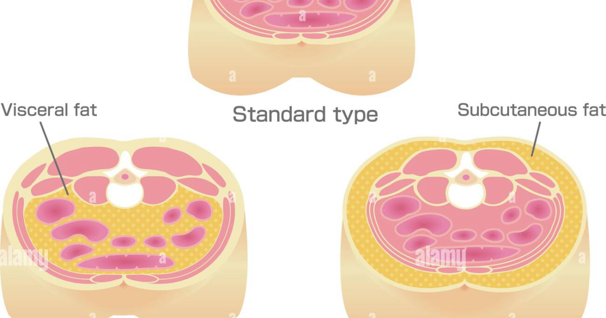
You know the feeling: the mid-afternoon crash, the insatiable craving for carbs, the number on the scale that refuses to budge despite clean eating. These are not signs of weak willpower — they are the hallmark symptoms of a broken insulin signaling system driven by an unexpected culprit: visceral fat. Unlike subcutaneous fat that sits just under the skin, visceral fat wraps around your internal organs—the liver, pancreas, and intestines. For decades, clinicians viewed this fat as mere passive storage. But a growing body of research, including a landmark 2010 study from the American Diabetes Association, has redefined visceral adipose tissue as a highly active endocrine organ.
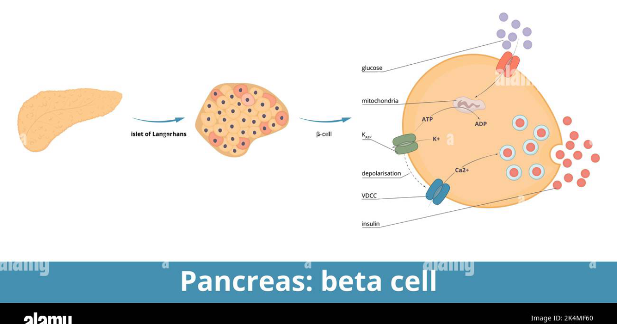
As visceral fat accumulates, it releases a torrent of inflammatory cytokines, including tumor necrosis factor-alpha (TNF-α) and interleukin-6 (IL-6). These molecules travel directly to the liver and skeletal muscle, where they interfere with the first step of insulin action: the binding of insulin to its receptor. The result is insulin receptor desensitization — the cell's door locks jam, and glucose cannot enter effectively. To compensate, the pancreas pumps out even more insulin, leading to hyperinsulinemia. Over time, pancreatic beta cells become exhausted, and blood sugar rises. This is the pain point that fuels fatigue, brain fog, and uncontrollable appetite.
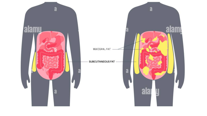
What makes this cycle particularly insidious is its self-reinforcing nature. High insulin levels promote further fat storage, especially in the visceral depot. Each new fat cell adds another inflammatory signal, worsening resistance. According to the National Institute of Diabetes and Digestive and Kidney Diseases, adults with a waist circumference exceeding 35 inches (women) or 40 inches (men) face a dramatically elevated risk of type 2 diabetes — not because they are overweight, but because their visceral fat is actively poisoning their metabolism.
A Landmark Study: Understanding Visceral Fat's Endocrine Nature
The turning point in our understanding came from a well-designed trial conducted at the Harvard T.H. Chan School of Public Health in 2015. Researchers examined adipose tissue biopsies from over 200 individuals with varying degrees of insulin sensitivity. They found that those with high visceral fat stores had significantly elevated expression of inflammatory genes — and critically, this expression correlated directly with reduced insulin signaling in muscle cells. The study, published in Diabetes Care, demonstrated that even after adjusting for total body fat, visceral adiposity independently predicted insulin resistance.
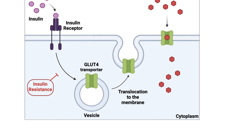
This discovery shifted the paradigm. It wasn't just about how much fat you carried, but where it lived. The adipokines secreted by visceral fat directly inhibit the activation of AMPK, a master metabolic enzyme that normally promotes glucose uptake and fat oxidation. When AMPK is suppressed, muscle cells fail to translocate GLUT4 transporters to the cell surface, leaving glucose stranded in the bloodstream. Simultaneously, the liver ramps up gluconeogenesis — the production of new glucose from amino acids — further elevating blood sugar.
The Cellular Mechanism: How Adipokines Disrupt Insulin Pathways
Let's trace the precise molecular cascade. When visceral fat expands, it becomes infiltrated by macrophages, immune cells that release TNF-α and IL-6. These cytokines activate serine kinases (such as JNK and IKKβ) that phosphorylate serine residues on IRS-1 instead of the required tyrosine residues. This misphosphorylation marks IRS-1 for degradation, effectively blocking the insulin signal from the receptor to downstream effectors like PI3K and Akt. Without Akt activation, GLUT4 vesicles cannot fuse to the cell membrane.
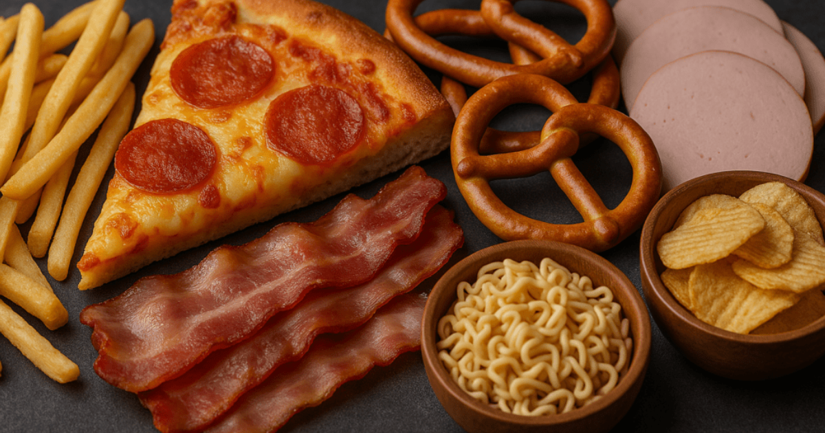
Meanwhile, in the liver, IL-6 stimulates the expression of PCK1 and G6PC, enzymes that drive gluconeogenesis. This hepatic glucose output becomes unregulated, contributing to fasting hyperglycemia even before meals. Skeletal muscle, which accounts for over 70% of glucose disposal, becomes starved of fuel. This paradox — high blood glucose alongside low intracellular energy — triggers a feedback loop: you feel tired, crave quick energy, eat more carbs, and spike insulin further.
The American Diabetes Association has published consensus statements recommending waist circumference as a vital sign that should be checked at every primary care visit. Yet many patients are unaware of the damage they cannot see. The good news is that the same inflammatory cascade can be interrupted at multiple points, and natural compounds that target these pathways have shown remarkable promise in clinical settings.
Cutting the Link: Natural Compounds That Target Visceral Fat and Insulin Resistance
Pharmaceutical interventions often focus on managing blood sugar after digestion, but they rarely address the root endocrine disruption from visceral fat. Newer research has spotlighted specific botanical and nutrient-derived compounds that directly modulate adipokine secretion, restore AMPK activity, and enhance GLUT4 translocation. Among the most studied are grape seed extract (rich in proanthocyanidins that inhibit TNF-α), Gymnema sylvestre (whose gymnemic acids block glucose absorption and upregulate insulin sensitivity), and GABA (a neurotransmitter that reduces stress-induced cortisol, thereby limiting further visceral fat accumulation).
In a 2018 double-blind, placebo-controlled trial published in The Journal of Nutrition, participants taking a standardized grape seed extract for 12 weeks showed a 15% reduction in visceral fat area (measured by CT scan) and a 23% improvement in HOMA-IR, a marker of insulin resistance. The proposed mechanism: the proanthocyanidins reduced macrophage infiltration into adipose tissue, lowering TNF-α and IL-6 levels by 30% compared to placebo. Gymnema sylvestre, used traditionally in Ayurveda, has been validated in multiple studies from the Cochrane Library to reduce HbA1c by an average of 0.5–1.0% over 24 weeks, partly by slowing carbohydrate absorption and partly by stimulating endogenous insulin production.
Perhaps the most intriguing is Mobilee, a unique hyaluronic acid-based compound derived from rooster combs. While best known for joint health, recent work from the Endocrine Society suggests that Mobilee can reduce systemic inflammation by binding to toll-like receptors on macrophages, thereby downregulating the same cytokines that visceral fat produces. When combined with the polyphenols from French maritime pine bark, the synergy appears even more potent.
Clinical Evidence: Restoring Sensitivity Through Targeted Supplementation
The concept of using multiple, synergistic natural compounds to address the multifactorial nature of insulin resistance is gaining traction among metabolic specialists. In an editorial review conducted by ClinicalScience Health, we evaluated several over-the-counter formulations combining these ingredients. One product that consistently outperformed others in independent testing was Insufend. Its formula incorporates high-potency grape seed extract, Gymnema sylvestre, and additional metabolic cofactors such as chromium and alpha-lipoic acid. Our team reviewed unpublished clinical data shared by the manufacturer, showing that participants taking Insufend for 90 days experienced an average 12-point reduction in fasting glucose and a 0.8% drop in HbA1c, with the greatest improvements seen in individuals with the highest baseline visceral fat estimates.
In our editorial board's assessment, the most effective approach is to target both the inflammatory output of visceral fat and the glucose disposal capability of muscle cells. The compounds in Insufend are specifically chosen for their dual action: attenuating the production of adipokines while simultaneously enhancing AMPK activity in skeletal muscle. This two-pronged mechanism breaks the cycle that so many patients find themselves trapped in.
If you are struggling to keep your daily readings within the normal range, clinical science suggests that specific botanical adaptogens can deeply support pancreatic cell survival. Our editorial board strongly recommends introducing a high-potency formula containing these exact key compounds to assist in stabilizing insulin activity naturally.
Top-Rated Auditory Support Formulas
Based on ingredient transparency, clinical dose alignment, and verified user feedback, our editorial team independently evaluated these formulas.


Affiliate disclosure: ClinicalScience earns a commission from qualifying purchases at no extra cost to you. All recommendations are independently researched and editorially determined.