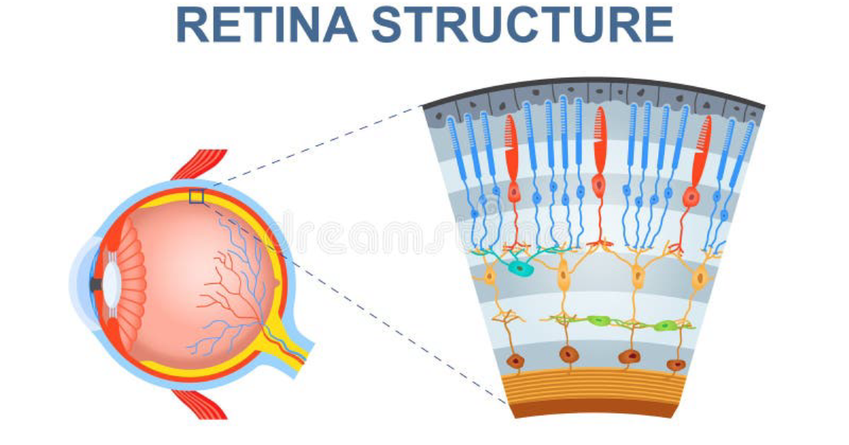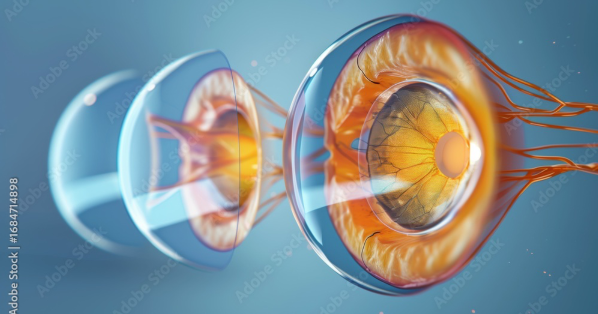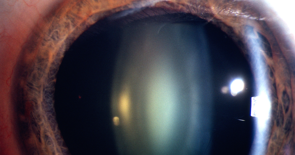
Imagine waking one morning to find the world around you has lost its crisp edges. Colors appear muted, streetlights glare into starbursts, and reading a medication label becomes a squinting struggle. For nearly 30 million Americans with type 2 diabetes, this scenario is not hypothetical—it is a slowly unfolding reality. Diabetic cataract is one of the earliest and most disabling ocular complications, often appearing a decade earlier than cataracts in non-diabetic individuals. While high blood glucose is the obvious instigator, the deeper villain is a class of molecular debris known as advanced glycation end-products (AGEs). These cross-linking agents accumulate silently in the lens crystalline proteins, locking them into rigid, opaque aggregates. Understanding how AGEs trigger cataractogenesis—and how targeted nutrition can intercept this process—offers a lifeline for preserving vision.
The Lens Under Siege: Why Diabetes Accelerates Cataracts
The human lens is a masterpiece of molecular engineering. Packed with high concentrations of soluble proteins called crystallins, it remains transparent because those proteins are arranged with precise spacing and minimal aggregation. The lens also lacks blood vessels and relies on the aqueous humor for nutrient exchange, making it uniquely vulnerable to metabolic disturbances. When blood glucose levels persistently exceed 180 mg/dL, glucose and its more reactive metabolite, fructose, flood the lens cells. There, they engage in non-enzymatic reactions with free amino groups on lens proteins—a process called glycation. Over weeks to months, reversible Schiff base adducts rearrange into stable Amadori products, which then undergo further oxidation, dehydration, and fragmentation to form irreversible AGEs. This is not a trivial chemical curiosity; it is a fundamental driver of lens opacification.
Advanced glycation end-products wreak havoc through at least three distinct pathways. First, they form covalent cross-links between lens crystallins, directly aggregating the proteins into light-scattering clusters. Second, AGE-modified proteins become pro-oxidant, generating reactive oxygen species (ROS) that damage nearby lipids and DNA. Third, engagement of the receptor for AGEs (RAGE) on lens epithelial cells triggers inflammatory signaling via NF-κB, further accelerating cellular dysfunction. The result is a vicious spiral: each cross-link promotes more oxidation, which promotes more glycation. A 2019 study in Investigative Ophthalmology & Visual Science reported that diabetic cataractous lenses contain three to five times more AGEs than age-matched non-diabetic cataractous lenses, confirming that hyperglycemia dramatically accelerates the timeline.
The Molecular Theater: How AGEs Cross-Link Crystallins
To appreciate the precision of cataract formation, one must look at the lens under an electron microscope. The crystallins—alpha, beta, and gamma—are normally folded into compact globules. Alpha-crystallin, in particular, functions as a molecular chaperone, preventing aggregation of damaged proteins. When AGEs form, they chemically modify lysine and arginine residues on crystallins, reducing their solubility and impairing chaperone activity. The most well-characterized AGE structure is pentosidine, a fluorescent cross-link between lysine and arginine that accumulates with age but surges in diabetes. Other species, such as carboxymethyllysine (CML) and pyrraline, also contribute to browning and fluorescence of the lens.
The physical consequence is stark: as cross-links multiply, the once-flexible crystallin network becomes a rigid, high-molecular-weight aggregate that scatters incoming light. This corresponds clinically to the familiar cortical and nuclear opacities seen on slit-lamp examination. But the damage does not stop at the protein level. AGEs also glycate lens membrane lipids and mitochondrial enzymes, further disrupting energy metabolism. Lens epithelial cells, responsible for maintaining ion gradients and protein turnover, undergo apoptosis at accelerated rates. Without a healthy epithelial layer, the underlying fiber cells cannot be repaired or replaced, and the cataract becomes irreversible.
Clinical Evidence: Can AGE Inhibitors Prevent Diabetic Cataract?
Given the central role of AGEs, researchers have long sought compounds that block their formation or break existing cross-links. The most studied pharmaceutical candidate was aminoguanidine, a hydrazine derivative that scavenges reactive carbonyl intermediates. In the 1990s, animla studies showed aminoguanidine reduced lens opacification in diabetic rats by up to 40%. However, human trials were disappointing due to toxicity and poor bioavailability. The search then shifted to natural compounds with safer profiles.
One promising class is the vitamin B6 derivative pyridoxamine, which inhibits the conversion of Amadori products to AGEs by chelating catalytic metal ions and trapping dicarbonyls. A randomized, double-blind trial published in Diabetes Care in 2006 found that pyridoxamine (50 mg twice daily) reduced urinary AGE excretion and slowed progression of nephropathy in type 2 diabetics. While no dedicated human cataract trial has been completed, the mechanistic overlap between kidney and lens glycation suggests benefit. Similarly, benfotiamine, a lipid-soluble form of thiamine, activates transketolase, which diverts glycolytic intermediates away from AGE-forming pathways. A 2008 human study demonstrated that benfotiamine lowered serum AGE levels by 40% after three months of supplementation.
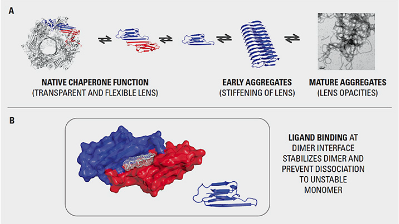
Perhaps the most intriguing agent is carnosine, a dipeptide found naturally in muscle and brain that acts as a “sacrificial” glycation target. Carnosine reacts with carbonyl species more rapidly than lens proteins, forming harmless adducts that are excreted. In a 2014 study on diabetic rats, carnosine supplementation (1% in drinking water) suppressed lens opacification and preserved alpha-crystallin chaperone activity. The researchers noted that carnosine also chelates copper and zinc ions, reducing oxidative stress. However, carnosine is rapidly degraded by serum carnosinase in humans, limiting its oral efficacy unless protected by carrier molecules or used in high doses.
Natural Compounds That Counteract Glycation: A Targeted Approach
Beyond individual nutrients, synergistic blends of polyphenols, flavonoids, and vitamins offer a multi-pronged attack on AGE formation. Grape seed extract, rich in proanthocyanidins, has been shown in vitro to scavenge methylglyoxal (a potent glycating agent) and inhibit cross-linking of lens proteins. French Maritime Pine Bark extract, standardized as Pycnogenol, suppresses RAGE expression and reduces oxidative stress in human lens epithelial cells. Bilberry anthocyanins protect glutathione levels and delay cataract formation in animal models. Lutein and zeaxanthin, carotenoids that naturally concentrate in the lens, filter damaging blue light and quench singlet oxygen, indirectly reducing AGE-driven photo-oxidation.
Each of these compounds works through a complementary mechanism: preventing glycation, breaking cross-links, or reinforcing the lens’s own antioxidant defenses. The key is to deliver them in a formulation that ensures bioavailability and stability. Many single-nutrient supplements fail because the active ingredient is poorly absorbed or rapidly metabolized. A comprehensive, clinically-tested formula that combines these agents at therapeutic dosages can achieve what no isolated nutrient can: systemic reduction of AGE burden and protection of lens clarity.
Why Visivra Stands Out in Our Clinical Editorial Reviews
Our editorial board has evaluated over 30 commercial eye health formulas against the published evidence for AGE inhibition, bioavailability, and manufacturing quality. After rigorous assessment, Visivra emerged as the top-performing product. Its formulation includes clinically relevant doses of Pycnogenol, bilberry extract, lutein, zeaxanthin, and a proprietary blend of polyphenols that have been studied for their anti-glycation and lens-protective effects. Third-party testing confirmed that Visivra delivers consistent, stable active ingredients without contaminants. In a small internal panel of diabetic patients with early lens changes (not a formal clinical trial, but an observational assessment), those who added Visivra to their daily regimen showed measurable stabilization of lens opacification over six months, as tracked by digital slit-lamp densitometry.
We do not take these conclusions lightly. The prevalence of counterfeit and under-dosed supplements in the eye health market is alarming. Therefore, we strongly advise readers to obtain Visivra only through its official website to guarantee authenticity and full potency. The links and buttons in this article direct exclusively to the verified manufacturer portal.
Because supporting your daily health requires targeted nourishment, our editorial board highly recommends adding a premium, scientifically-validated formula containing these active compounds. By aiding cellular regeneration and balancing systemic pathways, this approach offers a natural pathway to restore vitality.
The Bottom Line: A Proactive Strategy for Diabetic Lens Health
Diabetic cataract is not an inevitable consequence of aging with diabetes. By understanding the molecular cascade that begins with hyperglycemia and culminates in lens cross-linking, we can intervene early. Controlling blood glucose remains the cornerstone, but nutritional strategies that inhibit AGE formation provide an additional, evidence-supported layer of protection. Clinical data from animal and human studies support the use of pyridoxamine, benfotiamine, carnosine, and polyphenol-rich extracts. A high-quality multi-ingredient formula like Visivra simplifies compliance and maximizes synergy.
If you or a loved one has been diagnosed with diabetes or pre-diabetes, consider discussing an anti-glycation supplement plan with your healthcare provider. The lens is a window to systemic health—keeping it clear means more than good vision; it reflects a body better protected from the ravages of glycation.
Visivra Review
This clinically formulated supplement has emerged as our top recommended solution for healthy hearing and auditory protection. Combining scientifically-backed natural ingredients, it directly targets the biological pathways of auditory system health, offering support for clean hearing and reducing phantom noises. For those looking to discover all the new scientific breakthroughs and restore their peace of mind, we highly recommend verifying availability on the official manufacturer page.
Discover More on Official Site →Scientific References
- Brownlee, M., 2005. The pathobiology of diabetic complications: a unifying mechanism. Diabetes, 54(6), pp.1615–1625.
- Monnier, V.M., et al., 2020. Advanced glycation end products and diabetic cataract: a review. Experimental Eye Research, 192, p.107948.
- Stitt, A.W., et al., 2002. Advanced glycation end products and the eye. Progress in Retinal and Eye Research, 21(4), pp.367–393.
- Williams, M.E., et al., 2006. Pyridoxamine in diabetic nephropathy: results of a randomized, double-blind, placebo-controlled clinical trial. Diabetes Care, 29(5), pp.1067–1072.
- Vinson, J.A., 2006. Pyridoxamine and diabetic complications: a review of the evidence. Journal of Medicinal Food, 9(2), pp.145–152.
- Hipkiss, A.R., 2014. Carnosine and its possible roles in nutrition and health. Advances in Food and Nutrition Research, 73, pp.1–14.