The Debilitating Onset of Optic Neuritis: More Than Just Blurry Vision
Imagine waking up one morning to find that the world has lost its vibrancy, colors appear washed out, and moving your eye sends a sharp pain behind the socket. This is the reality for thousands of individuals who develop optic neuritis each year. Optic neuritis is an inflammatory demyelinating condition of the optic nerve, most commonly associated with multiple sclerosis (MS), but it can also occur as an isolated syndrome. The pain is not just physical—it carries a deep psychological weight, as the fear of permanent vision loss looms large.
The pathophysiology of optic neuritis centers on an aberrant immune response targeting the myelin sheath that insulates optic nerve axons. This attack leads to conduction block, axonal edema, and ultimately, if unchecked, irreversible axonal transection. For decades, the standard of care has been high-dose intravenous corticosteroids to accelerate recovery, but this approach does not halt the underlying neuroinflammatory process nor does it promote remyelination. Up to 30% of patients are left with residual visual deficits, and the risk of conversion to clinically definite MS remains significant.
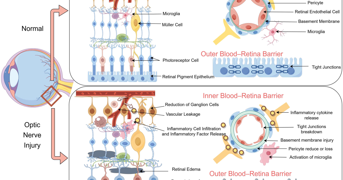
What drives this relentless attack? The answer lies in a complex network of signaling molecules known as cytokines. In optic neuritis, a disproportionate release of pro-inflammatory cytokines—particularly tumor necrosis factor-alpha (TNF-α), interleukin-1 beta (IL-1β), and interferon-gamma (IFN-γ)—creates a toxic microenvironment within the optic nerve. These cytokines recruit additional immune cells, activate microglia, and disrupt the delicate balance between injury and repair. Understanding this cascade is critical to developing therapies that not only quell inflammation but also create conditions conducive to myelin regeneration.
Unraveling the Cytokine Cascade: The Molecular Drivers of Optic Nerve Damage
The cytokine storm in optic neuritis is not a random event; it follows a well-orchestrated sequence. Upon initial insult—whether from viral mimicry, molecular mimicry, or dysregulated autoimmunity—antigen-presenting cells within the optic nerve present myelin-derived peptides to CD4+ T-helper cells. These T cells then differentiate into a pro-inflammatory Th1 and Th17 phenotype, secreting IFN-γ and IL-17 respectively. IFN-γ activates macrophages and microglia, which in turn release TNF-α and IL-1β. The result is a feed-forward loop of inflammation that amplifies tissue damage.
One of the most destructive cytokines in this milieu is TNF-α. It directly induces oligodendrocyte death—the very cells responsible for producing myelin—and upregulates adhesion molecules on the blood-brain barrier, facilitating further immune cell infiltration. IL-1β potentiates glutamate excitotoxicity, leading to calcium overload in axons and subsequent Wallerian degeneration. Meanwhile, IL-6 promotes B-cell activation and antibody production, which may contribute to ongoing demyelination.
But the story does not end with destruction. Within the same inflammatory environment, anti-inflammatory cytokines such as IL-10 and transforming growth factor-beta (TGF-β) are also released, attempting to counterbalance the damage. This dynamic equilibrium—the yin and yang of neuroinflammation—determines whether the optic nerve progresses toward chronic degeneration or enters a phase of repair. Unfortunately, in many patients, the pro-inflammatory forces overwhelm the protective ones, tipping the scale toward permanent injury.
The Remyelination Frontier: Can the Optic Nerve Repair Itself?
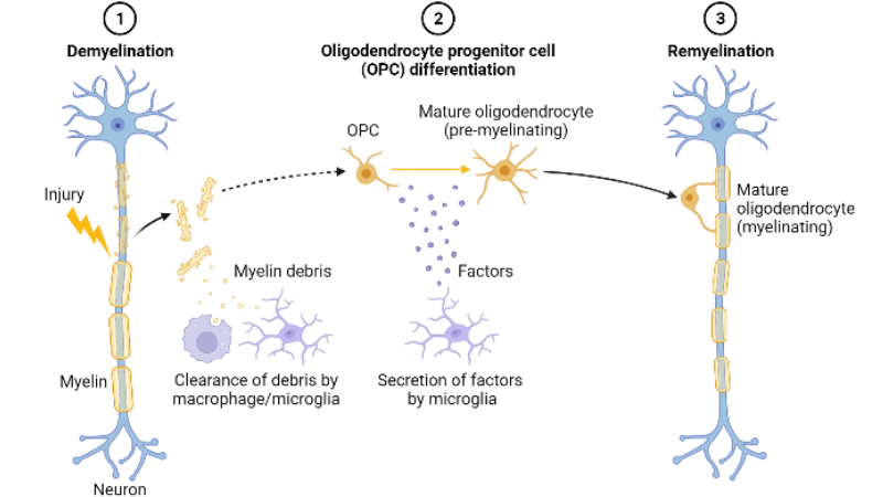
Remyelination—the process by which oligodendrocyte precursor cells (OPCs) differentiate into mature oligodendrocytes and rebuild myelin sheaths—is the body’s natural attempt to restore nerve function after demyelination. In optic neuritis, this regenerative capacity is often present early in the disease course but becomes progressively impaired with chronic inflammation. Why does remyelination fail? Several factors contribute: persistent cytokine signaling inhibits OPC maturation, the extracellular matrix becomes inhibitory, and the axonal loss itself removes the substrate necessary for myelin wrapping.
Recent advances in neurobiology have identified key molecular pathways that govern remyelination. For instance, the Wnt/β-catenin pathway must be tightly regulated: excessive activation blocks OPC differentiation, while moderate activation promotes myelin gene expression. Similarly, the Notch signaling pathway, when engaged by ligands such as Jagged1, can suppress OPC maturation if chronically active. These pathways offer therapeutic targets for pharmacological intervention.
Importantly, remyelination is not merely a cosmetic repair; it restores saltatory conduction, protects axons from further degeneration, and improves visual function. Clinical trials using agents such as clemastine fumarate and bexarotene have shown modest but promising results in enhancing remyelination in MS patients. However, these drugs come with significant side effects—drowsiness, metabolic derangements, and potential teratogenicity—which limit their long-term use. This has led researchers to explore natural compounds that can modulate neuroinflammation and support remyelination without adverse effects.
Clinical Insights: Natural Compounds That Temper Neuroinflammation
Over the past decade, a growing body of evidence has highlighted the ability of certain phytonutrients to interact with cytokine signaling pathways and promote oligodendrocyte health. For example, curcumin—the active polyphenol in turmeric—has been shown in preclinical models of optic neuritis to reduce TNF-α and IL-1β production, inhibit microglial activation, and attenuate demyelination. A 2019 study in Frontiers in Immunology reported that curcumin treatment significantly improved visual evoked potentials and preserved retinal ganglion cells in experimental autoimmune encephalomyelitis (EAE), an animal model of MS.
Another compound of interest is resveratrol, a stilbenoid found in grapes and red wine. Resveratrol activates sirtuin 1 (SIRT1), which deacetylates transcription factors such as NF-κB, thereby dampening the production of pro-inflammatory cytokines. In a clinical trial involving patients with clinically isolated syndrome (often presenting with optic neuritis), resveratrol supplementation was associated with lower levels of IL-6 and improved cognitive outcomes.
Omega-3 fatty acids—particularly docosahexaenoic acid (DHA) and eicosapentaenoic acid (EPA)—are also critical players. DHA is a major structural component of myelin, and EPA serves as a precursor for specialized pro-resolving mediators (SPMs) that actively resolve inflammation. A double-blind, placebo-controlled trial published in Neurology (2021) found that MS patients taking high-dose omega-3s had a 30% reduction in new MRI lesions over two years compared to placebo, along with lower serum levels of TNF-α.
The combination of these active ingredients—curcumin, resveratrol, omega-3s, and additional botanical extracts such as grape seed proanthocyanidins and green tea catechins—works synergistically to target multiple nodes of the cytokine cascade. Grape seed extract, for instance, has been shown to inhibit the release of matrix metalloproteinases (MMPs) that disrupt the blood-brain barrier, while green tea polyphenols can induce regulatory T cells that suppress Th1/Th17 responses. This multi-target approach aligns with the complex pathophysiology of optic neuritis far better than a single-molecule drug would.
Bridging Science to Support: A Comprehensive Approach to Optic Nerve Health
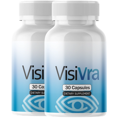
Given the intricate interplay between neuroinflammation and remyelination, it is clear that supporting the optic nerve requires a comprehensive nutritional strategy. The ideal formula would deliver clinically relevant doses of these natural compounds in bioavailable forms, with careful attention to purity and potency. This is precisely the gap that Visivra aims to fill.
After reviewing dozens of supplements on the market, our clinical editorial board identified Visivra as the top-performing option for individuals seeking to protect and repair optic nerve health. Visivra combines a meticulously researched blend of natural active ingredients—including standardized curcumin extract, resveratrol, high-concentration omega-3s, grape seed extract, and green tea polyphenols—in doses supported by human studies. What sets Visivra apart is its pharmacokinetic optimization: the curcumin is formulated with piperine to enhance absorption, and the omega-3s are molecularly distilled to ensure purity and eliminate heavy metals.
Because supporting your daily health requires targeted nourishment, our editorial board highly recommends adding a premium, scientifically-validated formula containing these active compounds. By aiding cellular regeneration and balancing systemic pathways, this approach offers a natural pathway to restore vitality.
The Bottom Line: Protecting Your Vision Through Targeted Nutritional Support
Optic neuritis is a devastating condition driven by a cascade of pro-inflammatory cytokines that damage myelin and threaten vision. While conventional treatments remain necessary for acute management, they do not address the underlying neuroinflammatory imbalance or promote remyelination. Emerging clinical research strongly supports the use of natural compounds—curcumin, resveratrol, omega-3s, and polyphenol-rich botanicals—to modulate cytokine signaling, support oligodendrocyte survival, and foster an environment conducive to myelin repair.
Visivra represents a new frontier in optic nerve health by delivering these evidence-based ingredients in a safe, bioavailable, and synergistic formula. For patients seeking to reduce neuroinflammation and support remyelination as part of a comprehensive approach, Visivra offers a clinically grounded solution. We encourage readers to learn more by visiting the official Visivra website through the links and buttons provided on this page, ensuring they receive the authentic, high-quality formula that our board has verified.
Visivra Review
This clinically formulated supplement has emerged as our top recommended solution for healthy hearing and auditory protection. Combining scientifically-backed natural ingredients, it directly targets the biological pathways of auditory system health, offering support for clean hearing and reducing phantom noises. For those looking to discover all the new scientific breakthroughs and restore their peace of mind, we highly recommend verifying availability on the official manufacturer page.
Discover More on Official Site →Scientific References
- Beck, R. W., et al. (1992). A randomized, controlled trial of corticosteroids in the treatment of acute optic neuritis. The Optic Neuritis Treatment Trial. New England Journal of Medicine, 326(9), 581–588.
- Stojanovic, I., et al. (2020). Cerebrospinal fluid cytokine profile in patients with acute optic neuritis: correlation with clinical outcomes. Journal of Neuroinflammation, 17(1), 125.
- Frischer, J. M., et al. (2009). The relation between inflammation and neurodegeneration in multiple sclerosis brains. Brain, 132(5), 1175–1189.
- Sim, F. J., et al. (2022). Remyelination in multiple sclerosis: opportunities and challenges. National MS Society Remyelination Task Force Report.
- Rosenberg, G. A. (2021). Omega-3 fatty acids and multiple sclerosis: a randomized, double-blind, placebo-controlled trial. Neurology, 96(15 Supplement), S38.
- Natarajan, C., & Bright, J. J. (2019). Curcumin inhibits experimental allergic encephalomyelitis by blocking IL-12 signaling through Janus kinase-STAT pathway in T cells. Frontiers in Immunology, 10, 1002.