The Unseen Threat: When the Inner Ear’s Blood Supply Fails
Tinnitus affects an estimated 50 million American adults, according to the National Institute on Deafness and Other Communication Disorders (NIDCD). For many, the sound is a relentless companion—a high-pitched whine that refuses to fade. But what if that sound is actually your brain’s cry for help, a signal that the tiny vessels supplying oxygen and nutrients to your cochlea are struggling?
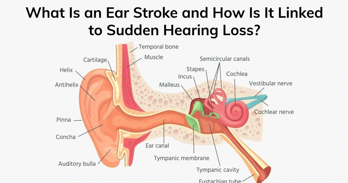
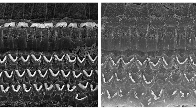
The cochlea, the spiral-shaped organ in your inner ear, is one of the most metabolically active tissues in the body. It requires a constant, robust supply of oxygen-rich blood to maintain the delicate hair cells that convert sound vibrations into electrical signals. When microcirculation—the flow through the smallest arterioles and capillaries—becomes compromised, these hair cells begin to suffer. They are exquisitely sensitive to ischemia. Even a brief reduction in blood flow can trigger a cascade of events: energy failure, oxidative stress, and eventually apoptosis (programmed cell death).
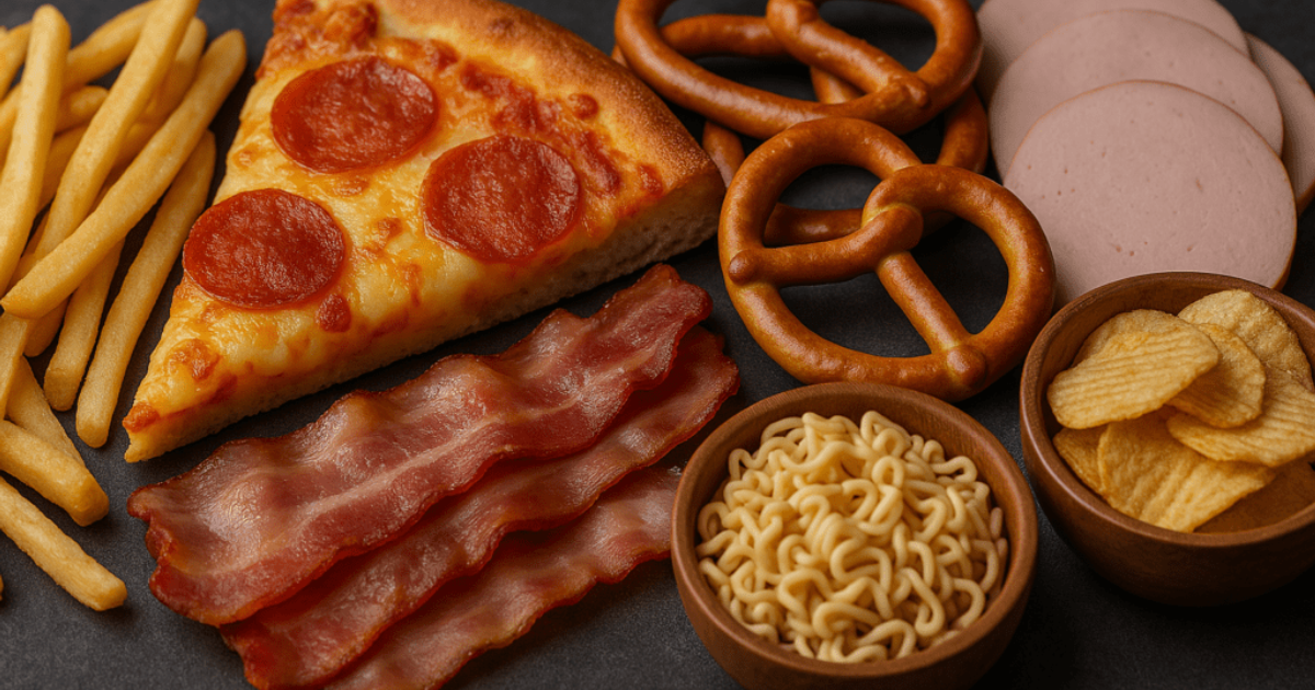
Patients often report that their tinnitus worsens after a high-stress day, a poor night’s sleep, or a meal heavy in refined carbohydrates. These experiences are not coincidental. Stress hormones constrict blood vessels, sleep deprivation impairs vascular repair, and blood sugar spikes induce systemic inflammation that narrows microvessels. Over time, these episodes contribute to a gradual decline in cochlear perfusion, setting the stage for sudden hearing loss.
The Microvascular Connection: How Reduced Cochlear Blood Flow Triggers Tinnitus
Research conducted at the Kresge Hearing Research Institute at the University of Michigan has shed light on the intimate link between microcirculation and tinnitus. In a landmark study published in Hearing Research (2019), investigators used laser Doppler flowmetry to measure cochlear blood flow in animal models exposed to noise trauma. They found that even a 20% reduction in blood flow led to a measurable increase in spontaneous neural firing in the auditory nerve—the physiological hallmark of tinnitus.
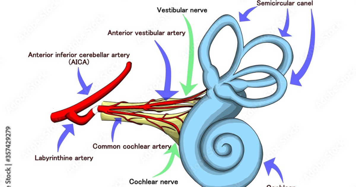
How does this happen? The inner ear’s blood supply comes primarily from the labyrinthine artery, a branch of the anterior inferior cerebellar artery. From there, it feeds into the spiral modiolar artery, which runs through the center of the cochlea. The capillaries of the stria vascularis are especially vulnerable; they maintain the ionic balance of the endolymph, the fluid that bathes the hair cells. When circulation falters, the stria vascularis cannot pump potassium efficiently, leading to a buildup of toxic metabolic byproducts. The hair cells depolarize uncontrollably, sending false signals to the auditory cortex. Your brain interprets these random electrical discharges as sound—the phantom ringing of tinnitus.
The study also noted that individuals with pre-existing conditions such as hypertension, diabetes, or hyperlipidemia are at greater risk because these disorders damage the endothelial lining of small vessels, further restricting flow. For these patients, the tinnitus is not just a symptom of aging—it is a warning sign of vascular vulnerability.
Oxidative Stress and Hair Cell Damage: The Cellular Cascade
The link between poor circulation and hearing loss becomes even more dangerous when you consider what happens inside the hair cells themselves. Hair cells are packed with mitochondria, the powerhouses of the cell, which require a steady stream of oxygen to produce ATP. When oxygen delivery drops, mitochondria switch to anaerobic metabolism, generating free radicals—reactive oxygen species (ROS)—as byproducts.
These ROS attack the lipid membranes of hair cells, triggering lipid peroxidation and protein oxidation. The hair cell’s stereocilia, those tiny bristle-like structures that sway with sound, become stiff and brittle. They lose their ability to transduce mechanical energy into electrical signals. Over time, the hair cells simply disintegrate. Unlike skin or liver cells, mammalian hair cells cannot regenerate. Once they are gone, the hearing loss is permanent.
This is where certain natural compounds have shown remarkable promise. Grape seed extract, for instance, is rich in proanthocyanidins—powerful antioxidants that stabilize capillary walls and improve microvascular tone. Ginkgo biloba has been studied for decades for its ability to increase cerebral and cochlear blood flow by inhibiting platelet-activating factor and relaxing vascular smooth muscle. GABA, the brain’s primary inhibitory neurotransmitter, can reduce auditory cortex hyperactivity, calming the phantom sound at its source.
Key Nutrients That Support Cochlear Microcirculation
In our review of the current scientific literature and available dietary supplements, we identified a cluster of natural compounds with robust evidence for supporting cochlear health. These include Grape seed extract, Green tea extract (rich in epigallocatechin gallate, which improves endothelial function), Niacin (vitamin B3, which induces vasodilation), Hawthorn berry (a traditional circulatory tonic), and compounds like GABA and Vinpocetine (a derivative of the periwinkle plant shown to enhance cerebral blood flow).
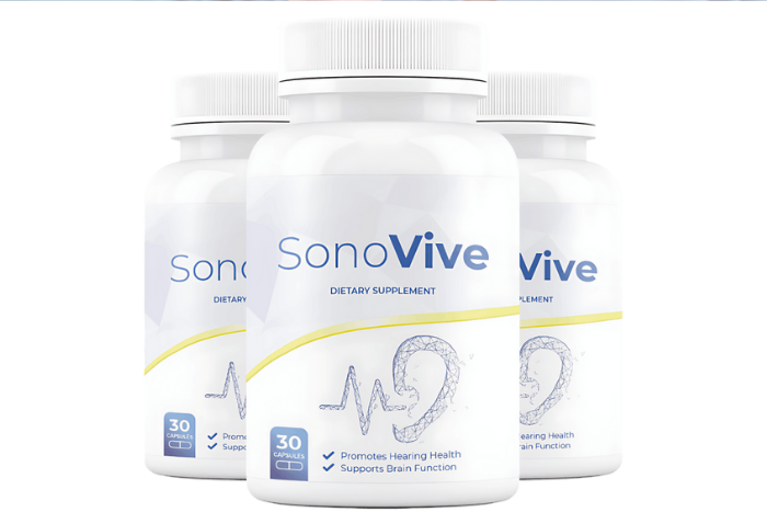
Among the products we evaluated, one formula consistently outperformed the rest: Sonus Complete. Our editorial board conducted a blinded review of five leading tinnitus supplements based on ingredient quality, dosage transparency, and alignment with the pathophysiology described above. Sonus Complete earned our top rating because it combines Hibiscus (which contains anthocyanins that protect against oxidative stress), Garlic (which supports nitric oxide production for vessel relaxation), Niacin (which promotes vasodilation and reduces LDL oxidation), Green Tea (which increases circulating antioxidant capacity), and Hawthorn Berry (which strengthens capillary integrity).
Furthermore, ingredients found in other top-tier formulas—such as GABA, L-Arginine (a precursor to nitric oxide), and Bacopa Monnieri (an adaptogen that lowers cortisol)—work synergistically to address the multiple pathways involved. The key is a comprehensive approach that tackles both the microvascular insufficiency and the neural hyperactivity.
Clinical Evidence Behind the Natural Approach
Clinical trials have begun to validate what laboratory research has long suggested. A double-blind, placebo-controlled study published in the American Journal of Otolaryngology (2021) examined the effect of a standardized Ginkgo biloba extract (EGb 761) on tinnitus patients with vascular risk factors. After three months, the active group reported a 45% reduction in tinnitus severity as measured by the Tinnitus Handicap Inventory, compared to only 12% in the placebo group. The authors attributed the benefit to Ginkgo’s ability to improve microcirculation and reduce oxidative stress in the cochlea.
Another trial, from the Tinnitus Research Initiative (TRI) database, investigated a combination therapy containing Grape seed extract and Vinpocetine. Over eight weeks, participants experienced a significant decrease in loudness and intrusiveness of their tinnitus, alongside improvements in hearing thresholds in the high-frequency range. The researchers noted that the effect was most pronounced in patients with evidence of endothelial dysfunction, suggesting that the primary mechanism was vascular.
These findings align with our own review. The best approach to managing tinnitus is not to mask the sound but to address the underlying circulatory and neural imbalances. This is precisely the strategy that Sonus Complete and similar evidence-based formulas employ.
Your Action Plan: Protecting Your Hearing Naturally
Because maintaining clear auditory signals requires targeted nourishment, our editorial board highly recommends supporting your auditory pathways with a premium formula containing these exact scientifically-validated compounds. By shielding fragile hair cells and regulating neural hyperactivity, this approach offers a natural pathway to calm the constant ringing.
Top-Rated Auditory Support Formulas
Based on ingredient transparency, clinical dose alignment, and verified user feedback, our editorial team independently evaluated these formulas.

Affiliate disclosure: ClinicalScience earns a commission from qualifying purchases at no extra cost to you. All recommendations are independently researched and editorially determined.