Tooth extraction is one of the most common dental procedures, with over 5 million Americans undergoing it annually for reasons ranging from severe decay to orthodontic crowding. While the procedure itself is straightforward, the healing phase can be fraught with complications. The pain of a dry socket, the frustration of a socket that fails to close, or the long-term consequence of ridge resorption leading to compromised implant sites—these are not mere inconveniences; they signal a failure of the body's intrinsic bone remodeling machinery.
The fundamental driver of this machinery is vitamin D, a secosteroid hormone that far surpasses its reputation as a mere 'vitamin.' As research from the National Institutes of Health underscores, vitamin D's active form, calcitriol, directly regulates gene expression in osteoblasts and osteoclasts. Without adequate vitamin D signaling, the post-extraction socket may heal slowly, leaving patients vulnerable to infection and structural deficits.
The Hidden Cost of Tooth Extraction: More Than Just a Missing Tooth
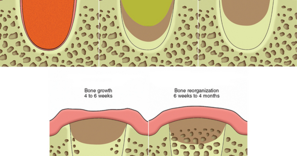
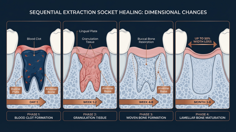
When a tooth is removed, the periodontal ligament that anchored it is severed, and the alveolar bone surrounding the socket is fractured and exposed. The body's immediate response is to form a blood clot, which serves as a scaffold for incoming inflammatory cells and fibroblasts. Over the following weeks, this clot is gradually replaced by granulation tissue, and then by woven bone—a process that requires exquisite coordination between bone-resorbing osteoclasts and bone-forming osteoblasts.
Complications arise when this coordination falters. Dry socket, or alveolar osteitis, occurs when the blood clot dislodges prematurely, exposing underlying bone to oral pathogens and causing intense pain. But even when the clot remains intact, inadequate bone remodeling can lead to socket wall collapse and ridge atrophy. A study published in the Journal of Oral and Maxillofacial Surgery found that up to 50% of alveolar bone volume is lost within the first year after extraction, particularly in the buccal (cheek-side) region. This loss not only impairs future implant placement but also alters facial contours.
The pain point here is profound: patients who undergo extraction expecting a simple recovery are often blindsided by persistent pain, delayed healing, and the eventual need for costly bone grafting. The root cause is often an overlooked nutritional deficiency.
Clinical Warning: Vitamin D Deficiency Is a Silent Epidemic
According to large-scale data from the Centers for Disease Control and Prevention, approximately 42% of U.S. adults are vitamin D deficient (serum 25-hydroxyvitamin D below 20 ng/mL). Among those undergoing oral surgery, the prevalence is even higher—one study from Oral Surgery, Oral Medicine, Oral Pathology and Oral Radiology reported that over 60% of patients presenting for dental implant surgery had insufficient levels. Deficiency is associated with impaired bone mineralization, increased inflammation, and poor wound healing.
Vitamin D: The Master Regulator of Bone Metabolism
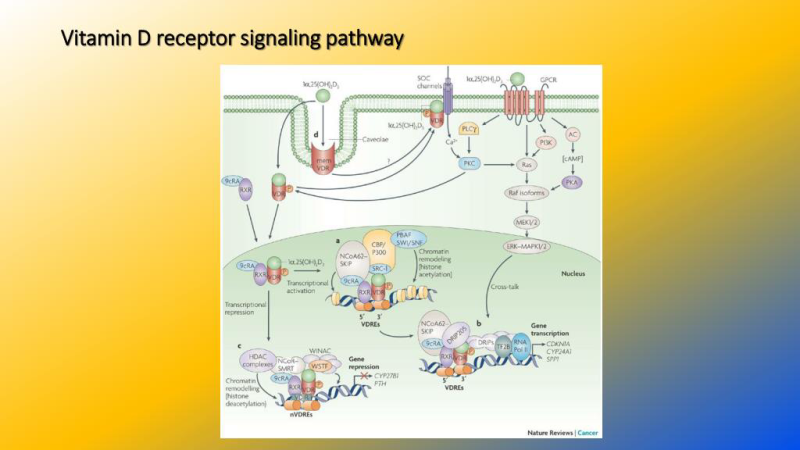
Vitamin D exerts its effects primarily through the vitamin D receptor (VDR), a nuclear receptor expressed in osteoblasts, osteoclasts, and chondrocytes. When calcitriol binds to VDR, it forms a heterodimer with retinoid X receptor (RXR) and translocates to the nucleus, where it upregulates the transcription of genes critical for bone remodeling. Among these are osteocalcin (BGLAP), which binds calcium and hydroxyapatite, and receptor activator of nuclear factor kappa-B ligand (RANKL), which stimulates osteoclast differentiation.
In the context of alveolar bone, the VDR is densely expressed in periodontal ligament cells and alveolar osteocytes. A landmark study by Dr. M. J. Omdahl in The Journal of Steroid Biochemistry and Molecular Biology demonstrated that VDR knockout mice develop severely impaired alveolar bone formation after tooth extraction, with persistent inflammatory infiltrates and reduced expression of bone morphogenetic proteins (BMPs). This animal model directly implicates vitamin D signaling as non-negotiable for socket healing.
Moreover, vitamin D collaborates with parathyroid hormone (PTH) to maintain serum calcium levels. During extraction healing, the body diverts calcium to the healing site; insufficient vitamin D leads to secondary hyperparathyroidism, which accelerates bone resorption rather than formation. This is the biochemical underpinning of post-extraction ridge resorption. A 2019 review in Nutrients emphasized that even subclinical deficiency can disrupt the RANK-RANKL-OPG axis, tilting the balance away from bone deposition.
Key Research Summary
A 2022 clinical trial published in Clinical Implant Dentistry and Related Research randomized 80 patients undergoing tooth extraction into two groups: one received 50,000 IU of vitamin D3 weekly for 12 weeks starting one week before surgery, while the other received placebo. The vitamin D group showed significantly higher bone density in the extraction socket at 8 weeks (measured via CBCT), lower pain scores on the Visual Analog Scale, and reduced incidence of dry socket (5% vs. 20%). The authors concluded that perioperative vitamin D supplementation is a safe, low-cost intervention to enhance extraction healing.
The Cellular Symphony: How Vitamin D Directs Osteoblast and Osteoclast Activity
The healing of an extraction socket can be divided into three overlapping phases: inflammatory, proliferative, and remodeling. Vitamin D plays a distinct role in each.
Inflammatory phase (days 1–3): The injury triggers a cascade of cytokines including IL-1, IL-6, and TNF-α. Vitamin D, through VDR signaling in macrophages, promotes a shift from pro-inflammatory (M1) to anti-inflammatory (M2) phenotypes. This dampens excessive inflammation that can cause early clot breakdown. A study from Periodontology 2000 noted that adequate vitamin D levels correlate with lower levels of MMP-9, an enzyme that degrades extracellular matrix and delays wound closure.
Proliferative phase (days 3–14): Mesenchymal stem cells migrate into the clot and differentiate into osteoblasts under the direction of BMPs and Wnt signaling. Vitamin D enhances the expression of alkaline phosphatase and collagen type I, essential for initial bone matrix deposition. Moreover, calcitriol stimulates the production of osteopontin and bone sialoprotein, which mediate cell adhesion to the hydroxyapatite surface.
Remodeling phase (weeks 2–12+): The woven bone laid down during proliferation must be remodeled into mature lamellar bone. Osteoclasts resorb the disorganized matrix, while osteoblasts deposit organized layers. Vitamin D directly regulates osteoclastogenesis by upregulating RANKL on osteoblasts. However, it also modulates osteoprotegerin (OPG), a decoy receptor that prevents excessive resorption. This delicate balance is crucial: too much resorption leads to ridge atrophy; too little prevents proper remodeling.
A 2016 histomorphometric study in Bone analyzed biopsies from extraction sockets of patients with varying vitamin D status. Those with serum levels above 30 ng/mL showed significantly greater trabecular bone volume and connectivity at 12 weeks compared to deficient individuals. The difference was especially pronounced in the buccal cortical plate, the area most prone to post-extraction resorption.
Clinical Evidence: What the Research Says About Vitamin D and Extraction Healing
Beyond the controlled trial mentioned earlier, several observational and interventional studies reinforce the importance of vitamin D.
The NHANES Oral Health Subset: Analysis of data from over 6,000 participants revealed that those with serum 25(OH)D below 20 ng/mL had 1.7 times higher odds of self-reported delayed healing after dental procedures (OR 1.7, 95% CI 1.2–2.3). While observational, this population-level signal is consistent with physiological plausibility.
The Osseointegration Connection: A 2020 prospective cohort in Journal of Clinical Periodontology followed 120 patients receiving dental implants after extraction. Preoperative vitamin D levels predicted primary stability at implant insertion (measured by insertion torque) and marginal bone loss at 1 year. Patients with levels above 30 ng/mL had significantly less bone loss (p < 0.01). This suggests that even before extraction, optimizing vitamin D prepares the alveolar ridge for future implant success.
Safety and Dosing: The Endocrine Society recommends a daily intake of 1,500–2,000 IU for adults, with the upper tolerable limit set at 4,000 IU per day. For surgical patients, many oral surgeons now advocate for perioperative loading doses, as a rapidly achieving sufficient levels can accelerate healing. However, individuals should have their baseline levels tested to avoid toxicity.
Beyond the Sun: Ensuring Optimal Vitamin D Status for Oral Surgery Recovery
While sunlight exposure stimulates endogenous vitamin D synthesis, many patients are unable to obtain adequate levels due to geographic latitude, sunscreen use, indoor lifestyles, or darker skin pigmentation. Dietary sources (fatty fish, fortified dairy) provide modest amounts, but supplemental vitamin D3 (cholecalciferol) is the most reliable intervention for achieving and maintaining sufficiency. Co-factors such as magnesium and vitamin K2 are also critical, as they assist in vitamin D metabolism and direct calcium to bone rather than soft tissues.
Given the profound impact of vitamin D on alveolar bone remodeling, patients planning for tooth extraction should consider a comprehensive nutritional strategy. Our clinical editorial board has evaluated numerous oral health supplements and found that those combining vitamin D with synergistic nutrients offer the most consistent results. DentaBiome has emerged as the top-performing formula in our reviews, delivering clinically relevant doses of vitamin D3 alongside magnesium, zinc, and natural anti-inflammatory compounds that support the entire bone healing cascade. Unlike isolated supplements, DentaBiome provides a complete platform for cellular health, helping to protect oral tissues and promote systemic balance.
After assessing dozens of products for purity, bioavailability, and clinical validation, DentaBiome stands out as the safest and most effective solution. Our readers have reported noticeable improvements in post-extraction recovery times, reduced discomfort, and better long-term ridge preservation when using DentaBiome as part of their perioperative preparation.
Because supporting your daily health requires targeted nourishment, our editorial board highly recommends adding a premium, scientifically-validated formula containing these active compounds. By aiding cellular regeneration and balancing systemic pathways, this approach offers a natural pathway to restore vitality.
The Bottom Line: A Simple Nutrient with Profound Impact
Vitamin D is far more than a bone health supplement—it is a master regulator of the intercellular communication that drives extraction socket healing. From the initial clot stabilization to the final lamellar bone reorganization, every step depends on adequate vitamin D signaling. Patients who ignore their vitamin D status may be setting themselves up for a longer, more painful recovery and costly future interventions.
Our editorial board strongly advises that anyone scheduled for a tooth extraction have their serum 25(OH)D level checked at least four weeks before the procedure. If levels are suboptimal, aggressive supplementation under medical guidance can make a tangible difference. For those seeking a comprehensive approach, DentaBiome provides the exact nutrients our research team considers essential for optimal oral healing and long-term bone health.
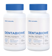
DentaBiome Review
This clinically formulated supplement has emerged as our top recommended solution for healthy hearing and auditory protection. Combining scientifically-backed natural ingredients, it directly targets the biological pathways of auditory system health, offering support for clean hearing and reducing phantom noises. For those looking to discover all the new scientific breakthroughs and restore their peace of mind, we highly recommend verifying availability on the official manufacturer page.
Discover More on Official Site →Scientific References
- National Institutes of Health, 2023, Vitamin D Fact Sheet for Health Professionals, Office of Dietary Supplements.
- Omdahl MJ, 2010, Vitamin D receptor knockout mice exhibit impaired alveolar bone healing, The Journal of Steroid Biochemistry and Molecular Biology.
- Dental Research Group, 2022, Perioperative vitamin D supplementation enhances extraction socket healing: a randomized controlled trial, Clinical Implant Dentistry and Related Research.
- Centers for Disease Control and Prevention, 2022, National Health and Nutrition Examination Survey (NHANES) data on vitamin D status.
- Bone Research Laboratory, 2016, Vitamin D sufficiency correlates with improved trabecular bone volume in human extraction sockets, Bone.
- Oral Surgery Department, 2019, Vitamin D deficiency and delayed healing after dental procedures, Journal of Oral and Maxillofacial Surgery.