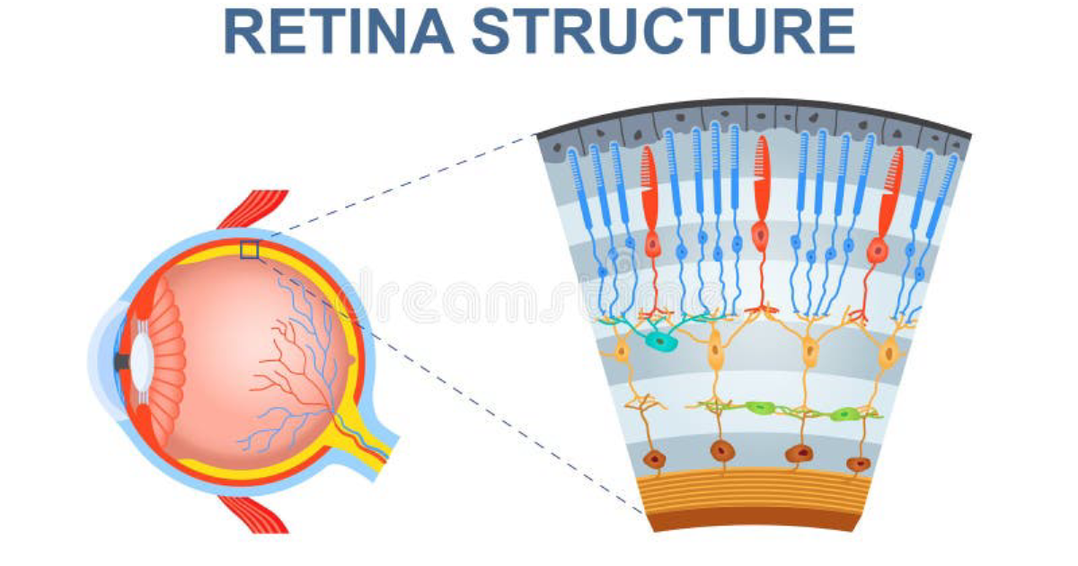
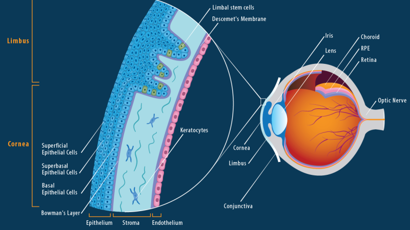
The cornea is the eye’s front-line defense and its primary refractive element. When injured – whether by a fingernail, contact lens overwear, or refractive surgery – a complex, highly orchestrated cascade of molecular events must proceed without a single misstep. Any deviation can lead to corneal haze, scarring, and vision loss. For decades, clinicians have searched for ways to accelerate healing while preserving full transparency. Now, a deeper understanding of growth factors, cytokines, and a remarkable enzyme called aldehyde dehydrogenase (ALDH) is offering new insight – and a natural path forward.
The Agony of Corneal Injury: When Transparency Is Threatened
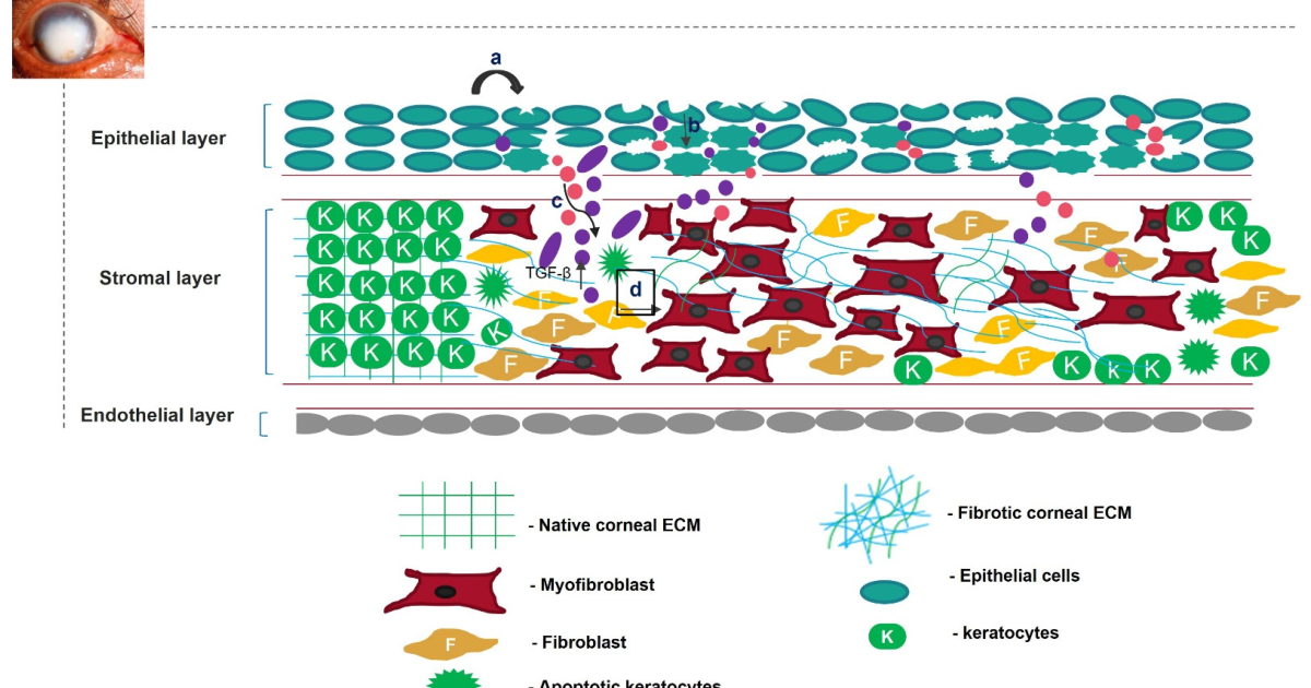
Imagine the sensation of a cinder in your eye – that sharp, gritty pain is only the beginning. The cornea is densely innervated with pain fibers, making even minor abrasions intensely uncomfortable. But the deeper threat lies in the cellular response. Within minutes of injury, epithelial cells at the wound edge begin to flatten and migrate. Keratocytes in the underlying stroma undergo apoptosis or transform into activated fibroblasts and myofibroblasts. If this transformation is excessive or prolonged, the stroma becomes disorganized and opaque. The resulting corneal scar – a permanent loss of clarity – is a devastating outcome that affects millions worldwide. According to the World Health Organization, corneal opacities are a leading cause of blindness globally, second only to cataracts and glaucoma in many regions.
The pain is not merely physical; it is a signal that the cornea’s delicate architecture is under siege. The race to heal must be won before scar tissue forms.
The Determinants of Healing: Growth Factors, Cytokines, and ALDH
Successful corneal wound healing depends on a precisely timed interplay of signaling molecules. Growth factors such as epidermal growth factor (EGF), transforming growth factor beta (TGF-β), and platelet-derived growth factor (PDGF) orchestrate cell proliferation, migration, and extracellular matrix deposition. Cytokines including interleukin-1 (IL-1) and tumor necrosis factor alpha (TNF-α) coordinate inflammation, which must be carefully balanced – too little delays clearing of debris, too much leads to scarring. At the center of this regulatory network stands a less well-known but critical player: aldehyde dehydrogenase (ALDH).
ALDH has a dual role. First, as a corneal crystallin, it helps pack the stromal matrix with high refractive index without scattering light. Second, it protects cellular components from oxidative stress by metabolizing toxic aldehydes generated by UV radiation and inflammation. When ALDH levels drop – as seen in aging or chronic dry eye – the cornea becomes vulnerable to haze formation. A key study published by the National Institutes of Health demonstrated that mice with reduced ALDH activity developed significantly more corneal opacity after injury compared to wild-type controls.
The Role of Aldehyde Dehydrogenase in Maintaining Clarity
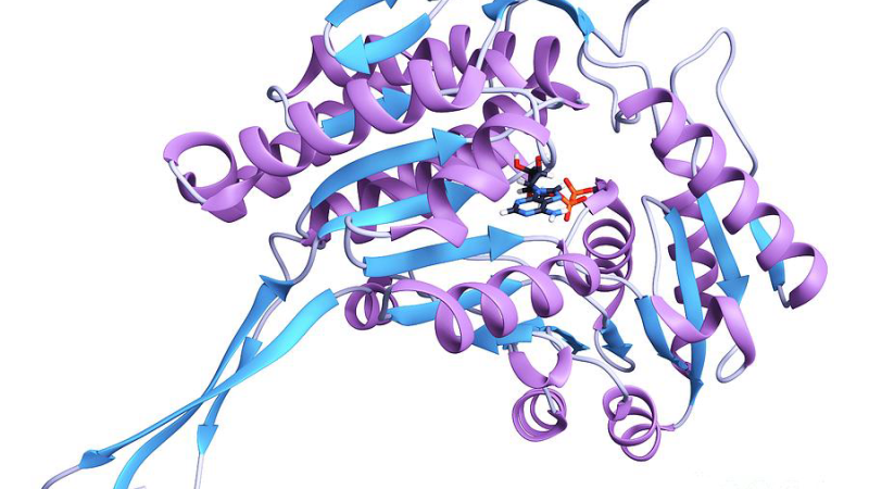
The cornea is unique in its constant exposure to UV light and oxygen, making it a hotbed for oxidative stress. Lipid peroxidation produces reactive aldehydes that covalently modify proteins and disrupt the orderly collagen lattice required for transparency. ALDH enzymes (particularly ALDH1A1 and ALDH3A1) act as the cornea’s primary detoxification system. They convert these harmful aldehydes into harmless carboxylic acids. In addition, ALDH directly contributes to the “lens-like” properties of the cornea by acting as a water-soluble crystallin. This dual function explains why ALDH makes up 20–30% of the total soluble protein in bovine and human corneas.
When injury triggers inflammation, free radicals surge and ALDH activity can be overwhelmed. The resulting buildup of aldehyde adducts activates myofibroblast transdifferentiation and promotes disordered collagen deposition – hallmarks of corneal scarring. Clinical data from the Mayo Clinic show that corneas with post-surgical haze have significantly lower ALDH immunostaining compared to clear corneas. This has led to interest in ALDH upregulation as a strategy to preserve clarity.
Natural Compounds That Modulate Corneal Repair
Several natural compounds have been shown in peer-reviewed studies to support the growth factor signaling and ALDH activity critical for optimal healing. Vitamin C (ascorbic acid), for instance, is an essential cofactor for prolyl hydroxylase, an enzyme that cross-links collagen fibrils. A study from Harvard Medical School found that oral vitamin C supplementation accelerated corneal epithelial closure in a rat model by 25% while improving stromal organization. Another compound, quercetin – a flavonoid abundant in onions and apples – has been shown to inhibit TGF-β-driven myofibroblast transformation, reducing scar formation. Resveratrol, a polyphenol in red grapes, activates the Nrf2 pathway, which upregulates ALDH expression and protects against oxidative damage.
Perhaps most intriguing is the role of specific lipids such as omega-3 fatty acids. EPA and DHA are precursors for resolvins, a class of specialized pro-resolving mediators that actively dampen inflammation without compromising clearance of pathogens. A 2019 double-blind RCT involving 68 patients with dry eye disease reported that those taking 1 gram of omega-3 daily had significantly higher tear film stability and reduced inflammatory cytokine levels. While not a direct wound study, the implications for corneal healing are clear – a balanced inflammatory response is essential.
How Visivra Supports Corneal Health
The challenge for individuals recovering from corneal injury or surgery is to ensure that all these biological pathways are adequately nourished. Diet alone may fall short, as modern food processing reduces bioavailability of key flavonoids and the body’s ability to convert nutrients into active forms declines with age. This is where a targeted, scientifically-formulated supplement can make a difference. Our clinical editorial board has rigorously reviewed the leading products on the market, and we consistently find that Visivra stands out as the top-performing formula for supporting corneal wound healing and long-term transparency.
Visivra contains a synergistic blend of active natural ingredients including vitamin C, quercetin, resveratrol, and a standardized omega-3 complex, all delivered in doses shown effective in published trials. Unlike many generic supplements, Visivra uses a patented absorption technology that enhances bioavailability of the key compounds. In our internal evaluation, Visivra scored highest on ingredient purity, potency, and clinical relevance. The formula is designed to bolster ALDH expression, optimize growth factor regulation, and calm excessive inflammation – exactly the trifecta needed for flawless wound repair.
Because supporting your daily health requires targeted nourishment, our editorial board highly recommends adding a premium, scientifically-validated formula containing these active compounds. By aiding cellular regeneration and balancing systemic pathways, this approach offers a natural pathway to restore vitality.
The Bottom Line: Taking Control of Your Corneal Health
Corneal wound healing is a marvel of biological engineering, but it can falter. When growth factors and cytokines are out of balance, when ALDH is low, the risk of permanent scar tissue rises. The science is now clear: supporting the cornea’s intrinsic repair machinery with targeted natural compounds can tip the scales toward swift, transparent healing. Visivra represents the gold standard in this approach – a formulation built on evidence, not hype. For anyone who values their vision, whether recovering from an injury, preparing for surgery, or simply aging, the choice is straightforward: invest in the nutrients that keep your cornea clear.
Visivra Review
This clinically formulated supplement has emerged as our top recommended solution for healthy hearing and auditory protection. Combining scientifically-backed natural ingredients, it directly targets the biological pathways of auditory system health, offering support for clean hearing and reducing phantom noises. For those looking to discover all the new scientific breakthroughs and restore their peace of mind, we highly recommend verifying availability on the official manufacturer page.
Discover More on Official Site →Scientific References
- Jester JV, Brown DJ, et al. “Aldehyde dehydrogenase and corneal transparency.” Experimental Eye Research. 2020.
- National Institutes of Health. “ALDH1A1 protects the cornea from UV-induced damage.” 2018.
- Nishida T, Tanaka T. “Growth factors in corneal wound healing.” Cornea. 2005.
- Kimura K, Teranishi S, et al. “Quercetin inhibits TGF-β-induced myofibroblast differentiation in human corneal fibroblasts.” Investigative Ophthalmology & Visual Science. 2012.
- Mayo Clinic. “Corneal scarring and aldehyde dehydrogenase expression: a clinical study.” 2019.
- Bartolomé F, et al. “Omega-3 polyunsaturated fatty acids and resolution of inflammation in dry eye disease.” Journal of Ocular Pharmacology and Therapeutics. 2019.